IHU Strasbourg at the Heart of MICCAI 2026: Awards, Innovations, and International Collaborations

IHU Strasbourg at the Heart of MICCAI 2026: Awards, Innovations, and International Collaborations

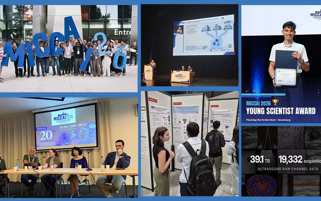
The 2026 edition of the MICCAI (Medical Image Computing and Computer-Assisted Intervention) international conference was a resounding success for our ecosystem. Following up on our first news update, we would like to highlight the scope of the achievements and awards earned by our teams and partners during that week in Strasbourg.

The scientific activity at IHU Strasbourg is based on a strong synergy between clinical practice, research, and the sharing of high-quality data. The results achieved at MICCAI 2026 perfectly illustrate the recognition of this work at the highest international level, with a total of 5 papers accepted for the main conference (out of more than 4,600 submissions, representing a highly selective acceptance rate of 27%).

A Hub for International Exchange

As the official sponsor of this year’s event, IHU Strasbourg has also established itself as a true focal point for the scientific community. The week was punctuated by major events alongside the main conference:

  • A pre-MICCAI event on Saturday. Coordinated by Prof. Caroline Essert, this event offered participants scientific presentations, a tour of the institute, and demonstrations on our experimental platform, led individually by Dr. Paolo Cabras, Jonas Mehtali, and the companies Scialytics and Iglobe Scientific.
  • A post-MICCAI seminar on Friday morning, featuring presentations by Dr. Yueming Jin (National University of Singapore—NUS) and Dr. Sharib Ali (University of Leeds).

A Prestigious “Young Scientist Award” for Liver Tumor Ablation Planning

One of the highlights of the conference was the presentation of the Young Scientist Award to Jonas Mehtali (funded by ITI HealthTech) for his work with Prof. Caroline Essert and Dr. Juan Manuel Verde on the planning of multi-needle thermal ablation.
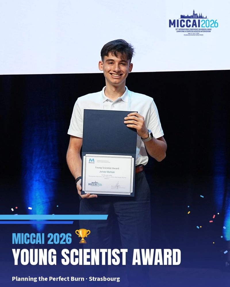
Photo credit: MICCAI 2026

Beyond the method developed at the ICube laboratory, this award highlights the clinical foundation provided by IHU Strasbourg: a unique, fully annotated, and documented liver dataset (50 cases, 94 tumors classified according to the COLLISION classification, 3D reconstructions, and reference ablation plans), compiled by Dr. Juan Manuel Verde and our teams. These data enabled a rigorous evaluation of the award-winning method, and the institute has published them in open access so that the scientific community can use them to develop and compare their own methods.

A Strong Scientific Presence: Publications and Spotlights

The publication activity involving IHU Strasbourg at this conference was once again remarkable:

  • The CAMMA team, led by Prof. Nicolas Padoy, demonstrated its dynamism with strong oral presentations and posters at the main conference and workshops. Among these successes, Julia Alekseenko, a Ph.D. student on the team (funded by the IHU’s DAIOR project), received a spotlight for her work on surgical phase recognition using hierarchical federated learning (Hi-FL). The event also highlighted the excellent contributions of Yaojun Hu and Ayush Gupta.
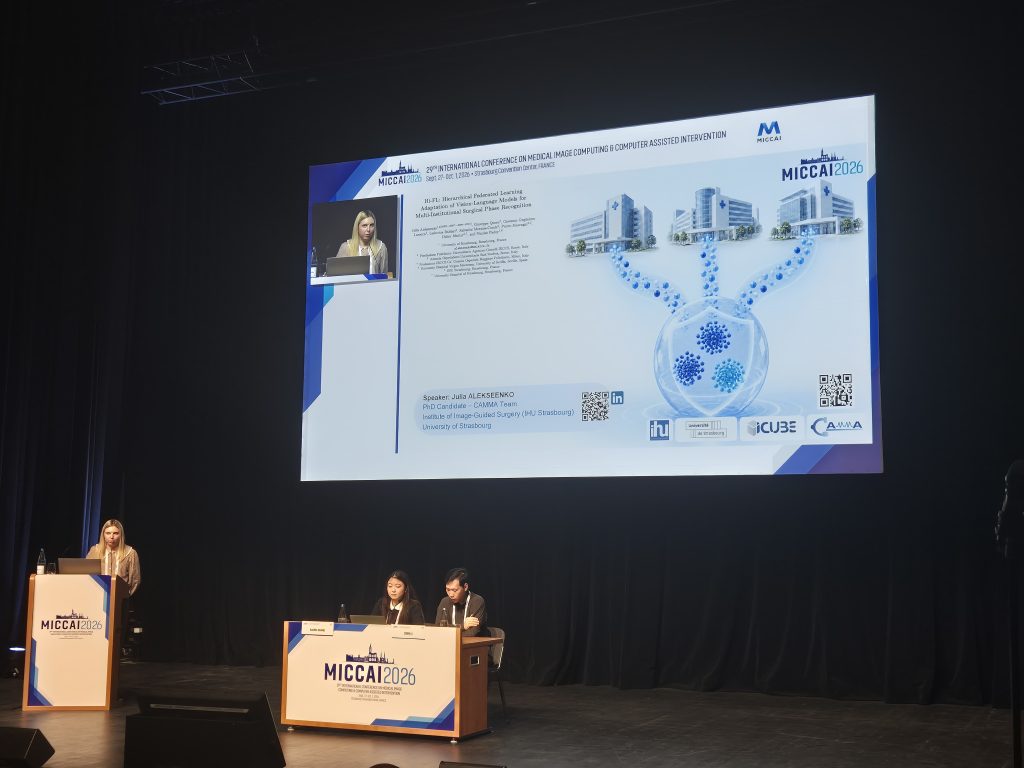
Photo Credit: MICCAI 2026

  • The Mimesis team (Inria / ICube), led by Dr. Stéphane Cotin, distinguished itself as part of the national MediTwin project with two notable papers (an oral presentation and a spotlight, by Francesco Dettori and Ilias Nahmed) on microwave ablation planning. This work draws directly on concepts developed at the institute and on in vivo preclinical data acquired entirely at the state-of-the-art experimental platform at IHU Strasbourg, during experiments on porcine models conducted by Dr. Juan Manuel Verde.

IHU at the Heart of Global Open Data With OpenH-RF
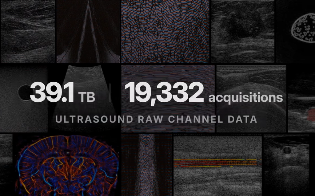
Photo credit: OpenH-RF

IHU’s influence is also evident in its contribution to OpenH-RF, a major international initiative led by Stanford University, Eindhoven University of Technology (TU/e), and NVIDIA. IHU Strasbourg is one of 38 contributing institutions (alongside Johns Hopkins, Siemens Healthineers, and the NIH) to what constitutes the largest open dataset of raw ultrasound signals ever published (hosted on the Hugging Face platform).

This project aims to train future foundation models for ultrasound. The contribution from IHU Strasbourg, led by Dr. Juan Manuel Verde, stems from its collaboration with the University of Basel (Prof. Philippe Cattin and Sidaty El Hadramy): a dataset of raw ultrasound images dedicated to bone imaging, which is now available to the community. The project was also the subject of a well-received presentation at the ASMUS workshop during MICCAI 2026.

Outreach and Institutional Roundtables

Finally, our teams’ expertise was called upon to enrich the discussions. In particular, Dr. Juan Manuel Verde was invited to serve as a panelist at the roundtable discussion celebrating the 20th anniversary of the AE-CAI workshop (’20 Years of AE-CAI: Looking Back and Looking Forward’), alongside eminent figures such as Prof. Nassir Navab, Prof. Stefanie Speidel, and Prof. Caroline Essert, moderated by Dr. Pierre Jannin.

Congratulations to all the clinicians, researchers, data scientists, and students who work every day to showcase Strasbourg’s university hospital research on the international stage!

Follow the IHU Strasbourg LinkedIn page to stay updated on our researchers’ latest breakthroughs.

Second Inter-IHU Day: Strengthening Collaboration and Sharing Expertise

Second Inter-IHU Day: Strengthening Collaboration and Sharing Expertise

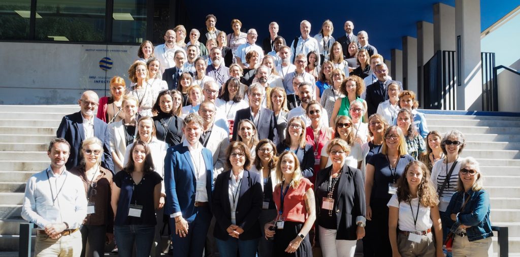
On September 22, Marseille hosted the second annual Inter-IHU Day. Co-organized by the IHU Méditerranée Infection and the IHU Prism, this national event brought together all members of the IHU-France alliance.
The shared goal? To create real momentum on the ground to promote the sharing of experiences and strengthen cooperation among the institutes.

The IHU-France Alliance: A Rapidly Expanding Network of Excellence

The creation of the University Hospital Institutes (IHU) marked a turning point for French medical research. The ambition was clear, and the approach was straightforward: to pioneer the medicine of tomorrow by bringing together scientific and medical excellence in a single location focused on a specialized field.

From just 6 in 2011, there are now 19, spread across the country and covering the full range of medical and surgical fields. This unique force brings together university hospitals, universities, and major public research organizations. It represents a “made in France” translational research capacity capable of competing with major international innovation hubs.

IHU-France Alliance Meeting During the Second Inter-IHU Day in Marseille

Working in Synergy to Accelerate Innovation in Health

The first Inter-IHU Day held in Paris laid the groundwork for the network; this edition in Marseille marked the transition to action.
Gathered for plenary sessions and several thematic workshops, about 100 participants discussed topics central to the IHUs’ missions: network dynamics and collaborations, prevention, data utilization, and training.
One major area of cooperation identified is the utilization of health data, with the IHU-France alliance playing a decisive role at the national level in facilitating the use of health data and developing reliable and sovereign AI models. In this regard, the flagship national MediTwin project—which develops digital twins for personalized medicine, led by Dassault Systèmes and bringing together seven IHUs with Inria—is a perfect example of the IHUs’ ability to collectively create a virtuous cycle that links healthcare to research, thereby providing solutions to unmet medical needs.

Interdisciplinarity Dedicated Exclusively to Patients

The joint organization of this event by the IHU Méditerranée Infection (infectious diseases) and the IHU Prism (oncology) perfectly illustrated interdisciplinarity—the hallmark of the IHU-France alliance and a true asset for innovation in healthcare.

Working to safeguard the common good of health, meet clinical needs, maintain the agility required for innovation, and bring together fields of expertise in research and care: this is the IHU-France Alliance’s formula for developing large-scale projects. Ultimately, this collective effort has a single guiding principle: to rapidly transform these scientific advances into concrete solutions for patients, both today and tomorrow.

Are you passionate about the integration of Artificial Intelligence and clinical research? Follow IHU Strasbourg on LinkedIn so you don’t miss any of our upcoming developments.

French Premiere at the IHU Strasbourg: Artificial Intelligence Aids in Pancreatic Diagnosis with the mAI Companion® Medical Device

French Premiere at the IHU Strasbourg: Artificial Intelligence Aids in Pancreatic Diagnosis with the mAI Companion® Medical Device

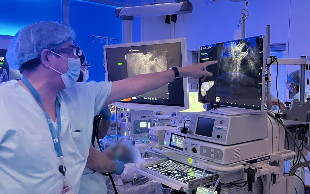
On August 25, 2026, the gastrointestinal endoscopy team at the Strasbourg University Hospitals (HUS) validated the first clinical use in France of the mAI Companion® device at IHU Strasbourg. Co-developed by iGlobe Scientific, a joint venture between IHU Strasbourg and Medi-Globe, this CE-marked medical device uses artificial intelligence to assist in endoscopic ultrasound examinations of the pancreas, particularly for the early detection of pancreatic cancer.

Inside the Operating Room: A Decisive Morning

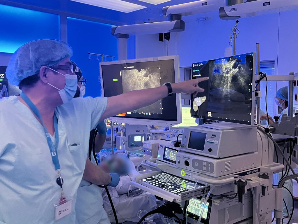
For this first procedure, four routine clinical examinations were performed. The procedure involved Prof. François Habersetzer, Dr. Pierre-Emmanuel Mayer, and Dr. Pierre-Yves Christmann from the HUS; Dr. Leonardo Sosa Valencia from the IHU Strasbourg; and Jean-Paul Mazellier, technical director of iGlobe Scientific.

Dr. Leonardo Sosa Valencia, an expert in endoscopic ultrasound at the IHU Strasbourg – Photo credit: iGlobe Scientific

Real-Time AI Assistance for Clinicians

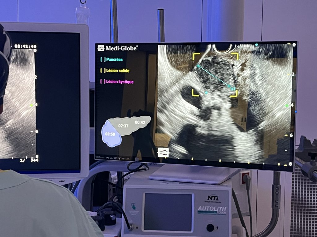
The mAI Companion® device connects easily to any type of endoscopic ultrasound equipment. It receives a copy of the video feed, which it analyzes locally and in real time, then displays annotations on a dedicated screen. This information helps the clinician examine the pancreas by drawing their attention to specific areas and enabling them to perform as thorough an analysis as possible. The system operates in a closed loop; no data is transmitted externally, ensuring absolute security of medical data. At all times, the clinician remains in control and retains full responsibility for interpretation and clinical decision-making.

mAI Companion® artificial intelligence (AI) assistant creating annotations on specific areas of the pancreas during the patient’s examination – Photo credit: iGlobe Scientific

The Success of a Franco-German Partnership in R&D and Technology Transfer

The mAI Companion® innovation exemplifies the transition from cutting-edge academic research to a functional clinical tool that benefits patients.

The story began in 2021 with the APEUS (Augmented Pancreatic Endoscopic Ultrasound) project, supported by the ARC Foundation. APEUS was led at the IHU Strasbourg jointly by Dr. Leonardo Sosa Valencia, a gastroenterologist specializing in endoscopic ultrasound, and Prof. Nicolas Padoy, director of the CAMMA (Surgical Data Science and Artificial Intelligence). The promising results obtained convinced the German company Medi-Globe to enter into a research and development collaboration with the IHU in April 2022.

This collaboration led to the creation of the joint venture iGlobe Scientific SAS, established in Strasbourg to develop the medical device, which is marketed by Medi-Globe GmbH, the device’s authorized manufacturer. Having obtained CE marking in early 2026, mAI Companion® demonstrates the strength of Strasbourg’s ecosystem for research and innovation in the field of medical technology.

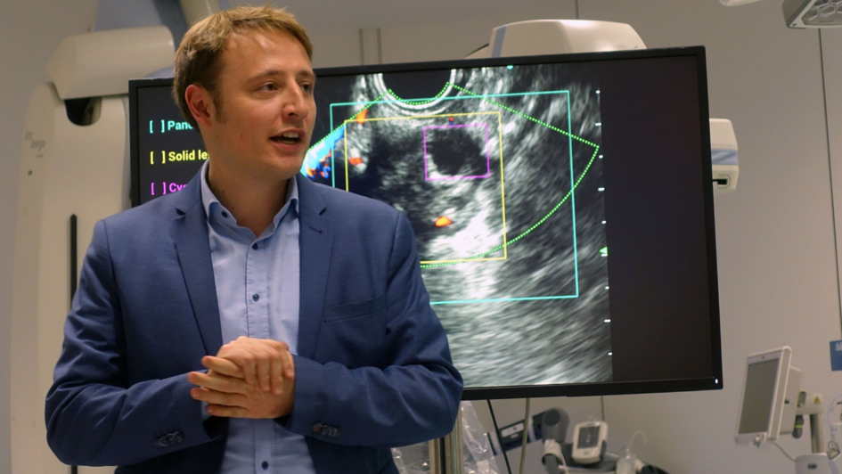
The AI assistant, mAI Companion®, complies with the European Medical Device Regulation (MDR) and will be CE-marked in 2026, with Dr. Markus Schönberger, Managing Director of Medi-Globe—Photo credit: IHU Strasbourg

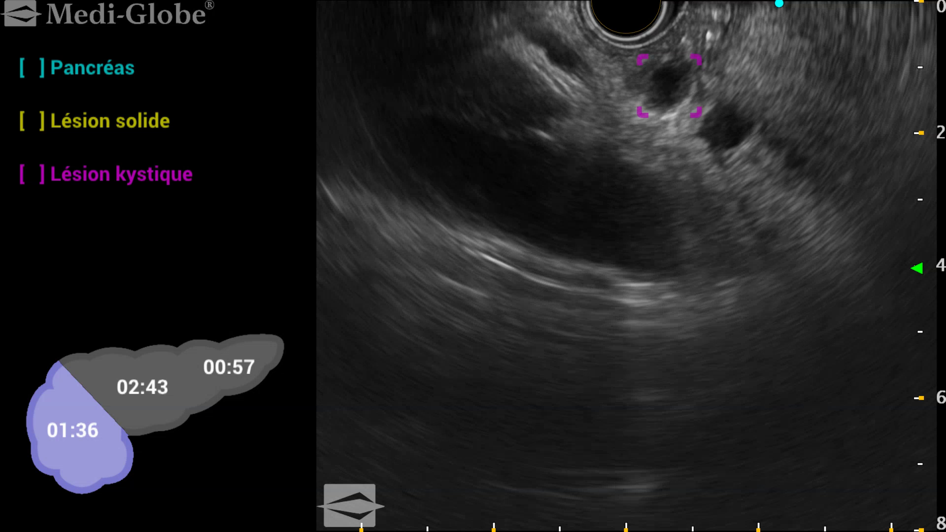
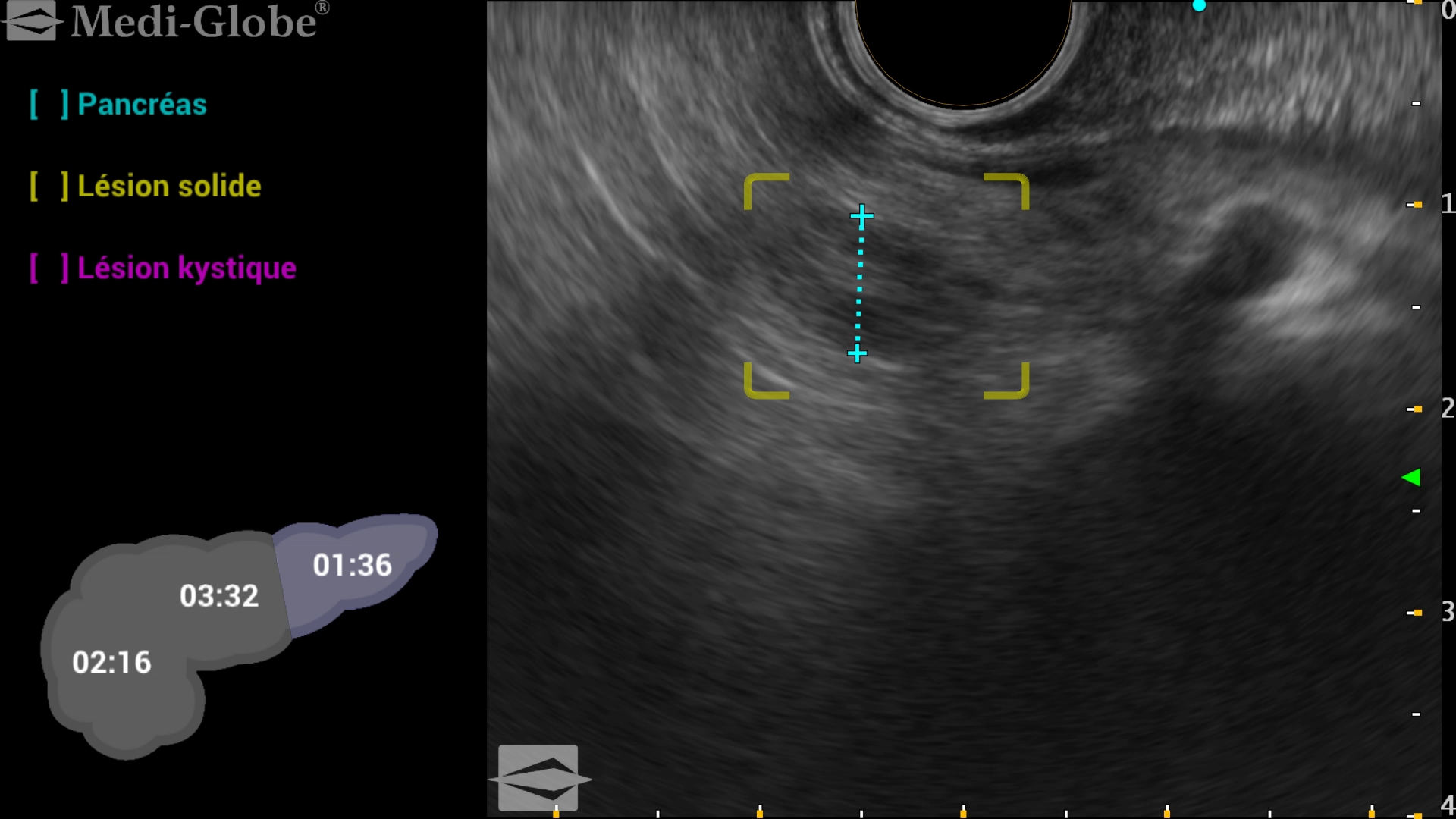
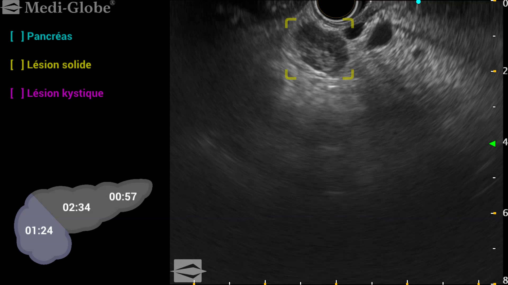
Examples of lesions detected by the mAI Companion® AI assistant

Cystic lesion of the head of the pancreas

Photo credit: iGlobe Scientific


Cystic lesion of the head of the pancreas of 6mm detected by mai companion during a pancreatic evaluation for pancreatitis, corresponding to a IPMN of branch duct with a benignant aspect.


Solid lesion of the tail of the pancreas

Photo credit: iGlobe Scientific


Solid lesion of the tail of the pancreas with less than 10mm, another IPMN with mucus inside detected by mai companion after acute pancreatitis that look solid due to the content.

Solid lesion of the pancreatic head

Photo credit: iGlobe Scientific


Solid lesion of the head of the pancreas between 10 to 15 mm, possible NET.

To learn more: Check out our retrospective on the CE marking of mAI Companion® and our article on the impact of AI on the detection of pancreatic lesions.

Are you a healthcare professional or an industry partner interested in learning more about integrating mAI Companion®?
Would you like to contact the iGlobe Scientific team?

Fondation ARC Les Hôpitaux Universitaires de Strasbourg (NHC) CAMMA (Computational Analysis and Modeling of Medical Activities) Université de Strasbourg iGlobe Scientific Medi-Globe
Precision Robotics for Hearing Surgery: IHU Strasbourg launches a New Program of Clinical Excellence and R&D

Precision Robotics for Hearing Surgery: IHU Strasbourg launches a New Program of Clinical Excellence and R&D

Medical innovation takes a major step forward in Alsace

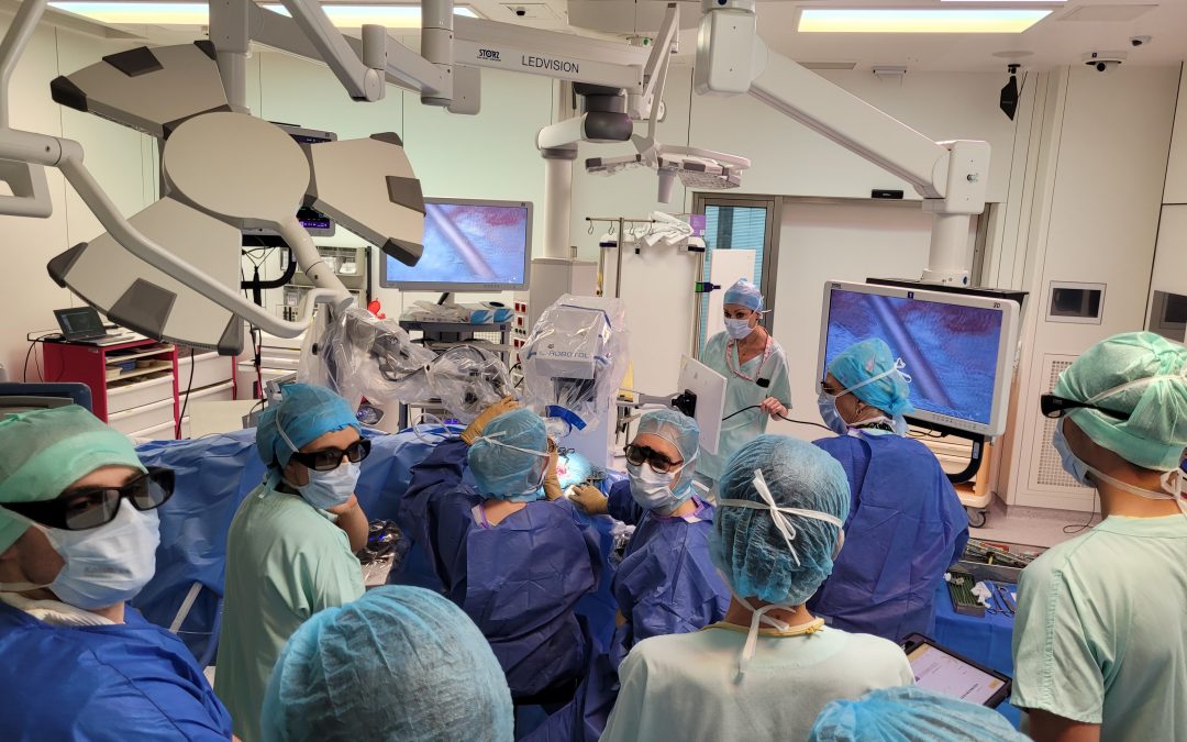
On July 16, 2026, the technical platform at the IHU Strasbourg hosted a landmark medical procedure: the implantation of a cochlear implant (for profound bilateral hearing loss) that simultaneously incorporated 3D exoscopy and otologic robotics.

A Leading Clinical and Industrial Collaboration

This complex procedure was successfully performed by the surgical team in the ENT department at the Strasbourg University Hospitals, under the direction of Prof. Christian Debry (director of the IHU and head of the ENT department), and led by Prof. Anne Charpiot and Dr. Carine Eyermann. The procedure on this first adult patient relied on a combination of two complementary, state-of-the-art technologies:

  • The VITOM® 3D Exoscope – Karl Storz: This high-definition visualization system projects a 3D image onto a dedicated surgical monitor. It provides optimal perception of depth and anatomical landmarks in the middle ear, improving ergonomic workflow compared to a traditional microscope.
  • The RobOtol® robot – Collin Medical: The first robotic system designed specifically for ear microsurgery, it assists the surgeon in the micrometric insertion of the implant electrode into the cochlea, thereby ensuring the safety of the procedure and maximizing the preservation of the delicate structures of the inner ear.

Spreading Technology and Preparing for the Future

While this dual technological approach has already been tested in other hospitals in France, its implementation at the IHU Strasbourg and the HUS is part of a larger initiative. For the IHU Strasbourg and its partners, the challenge does not end with this initial success. Drawing on the experience gained in the field, the teams are actively participating in the development and optimization of future robots designed for ENT and other precision medical specialties.

Prospects for Research and Training

Starting in September 2026, this clinical program will get into full swing with one to two scheduled outpatient procedures per month. The upcoming integration of the ENT and neurosurgery departments will round out the institute’s areas of expertise.

Beyond the immediate benefits for patients, this activity in the technical platform will serve as the foundation for new training programs for residents, specialists, and operating room nurses (IBODE). It will also fuel ambitious translational research projects in the fields of surgery and AI-enhanced imaging, fitting naturally into the roadmap for the IHU Strasbourg’s major new collaborative projects in close partnership with the HUS.

Learn more:

  • The ENT (Ear, Nose, and Throat) and Head and Neck Surgery Department at HUS: Click here
  • VITOM® 3D Exoscope (Karl Storz): Click here
  • RobOtol® robot (Collin Medical): Click here
HUS de Strasbourg
Karl Storz
Collin Medical
MediTwin 2026 Symposium: Advances in Virtual Twins for the Medical Practice of Tomorrow

MediTwin 2026 Symposium: Advances in Virtual Twins for the Medical Practice of Tomorrow

The Medicine of Tomorrow: The MediTwin Consortium Reviews Progress on Virtual Twins at Its Scientific Symposium

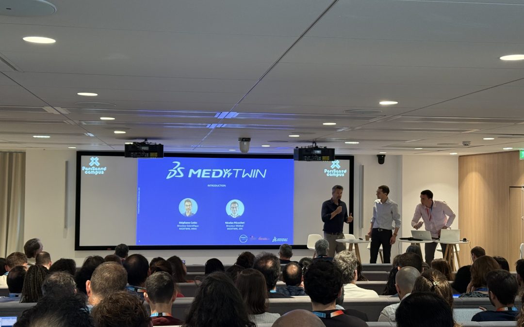
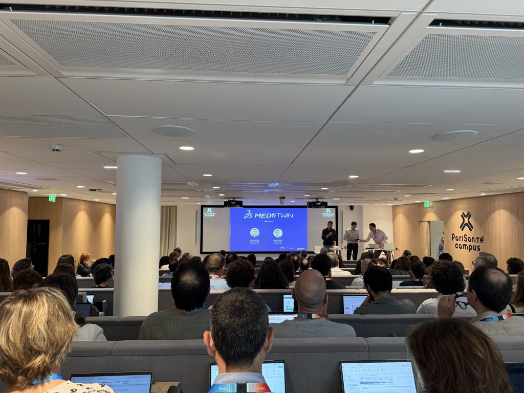
On June 3, PariSanté Campus hosted the symposium for the MediTwin project led by Dassault Systèmes to create virtual twins for the medicine of tomorrow. This event provided an opportunity for the consortium’s partners to present concrete progress in the translational and technological research being conducted.

The MediTwin consortium provides an update on the progress of virtual twins

The MediTwin Project

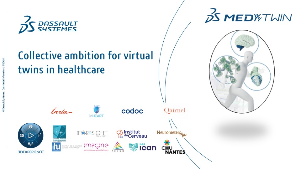
Launched with the ambition of revolutionizing medical practice, MediTwin develops personalized virtual twins for patients. These virtual replicas of organs or medical conditions make it possible to simulate treatments, predict disease progression, and ensure the safety of surgical or therapeutic procedures even before they are performed in a clinical setting.

Developed over five years (from 2024 to 2029), this large-scale initiative receives financial support from France 2030, affirming the national commitment to positioning the country as a leader in digital health.

A Consortium of Excellence

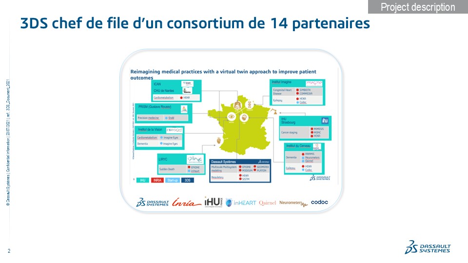
MediTwin’s strength lies in its multidisciplinary approach, bringing together a global industry leader—Dassault Systèmes—innovative startups—inHEART, Qairn, UsefulProgress, PredictMedical, and Codoc—a leading research institute—Inria— seven university hospitals: Foresight, ICAN, Imagine, Institut du Cerveau, Liryc, PRISM, and Strasbourg, and the Nantes University Hospital.

MediTwin: A Consortium of Excellence

A rich and collaborative program

The symposium began with a morning of plenary lectures focused on the project’s clinical applications in prevention and care within cardiology, oncology, and neurology.

MediTwin: A comprehensive and collaborative program

The afternoon was devoted to technical workshops and collaborative working groups. Researchers and clinicians were able to discuss image segmentation, multi-fidelity simulation, data assimilation, and the integration of artificial intelligence and numerical simulation. A poster session and presentations of Inria’s cutting-edge SOFA and Fed-BioMed software platforms rounded out this day of scientific exchange.

By bringing together government resources, the agility of startups, the rigor of academic research, and the expertise of clinicians, MediTwin is steadily fulfilling its promise: to deliver more predictive, personalized, and safe medicine for the direct benefit of patients.

Learn more about the MediTwin project:
www.3ds.com/fr/science/meditwin

RHU ENVISAGE: Revolutionize the treatment of heart valve diseases.

RHU ENVISAGE: Revolutionize the treatment of heart valve diseases.

RHU ENVISAGE: A medical, scientific, and technological alliance to transform the management of valvular heart disease.

Valvular heart disease refers to conditions affecting the heart’s valves that can lead to heart failure. Diagnosis relies primarily on echocardiography, which allows for the assessment of valve damage and the feasibility of interventions. The ENVISAGE (Enhanced Valves Interventions and Safe AI Generated End results) University-Hospital Research (RHU) program is developing a decision-making platform guided by echocardiography enhanced by artificial intelligence for optimized management of valvular heart disease. Led by the Bordeaux University Hospital, the ENVISAGE consortium brings together leading university hospital and industry partners around a common goal: to develop an AI solution that is easy to use in clinical practice to guide patient selection, planning, and the prediction of outcomes for transcatheter interventions.

RHU ENVISAGE

Photo credit: RHU ENVISAGE

The IHU Strasbourg and the MOSaiC platform

The IHU Strasbourg contributes to the ENVISAGE program with its expertise and its online collaborative platform, MOSaiC, designed for the annotation and evaluation of medical videos. MOSaiC was developed by Prof. Nicolas Padoy and his CAMMA (Computational Analysis and Modeling of Medical Activities) team to accelerate data science projects in surgery and interventional medicine. The IHU Strasbourg’s involvement in ENVISAGE demonstrates its commitment to providing powerful tools to accelerate the transfer of translational research findings to patients.

Want to learn more?

Presentation video: www.youtube.com/watch?v=3rJZB5DkOiM
Project website: www.envisage-rhu.com

Do you have a question, an idea, or need more information?
www.envisage-rhu.com/contact

Project partners

Partenaires du projet RHU Envisage